Handy FluorCam
Chlorophyll Fluorescence Imaging from Individual Cells to Plant Populations

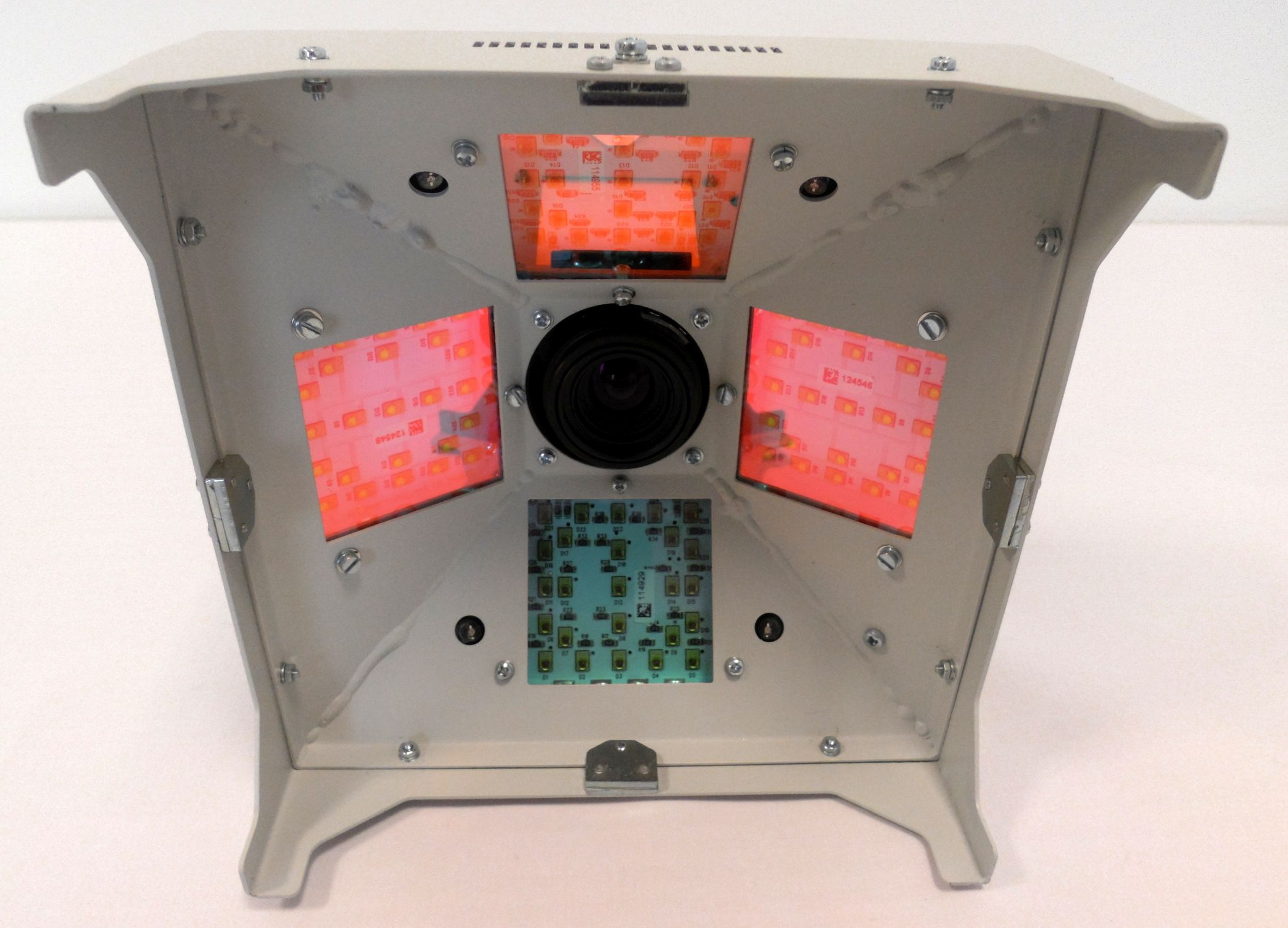
Handy FluorCam FC 1000-H is a lightweight, portable device designed for time resolved chlorophyll fluorescence imaging of leaves, small plants, leaf segments, mosses, lichens, seeds, roots, tissues on plates or algal colonies both in a field and laboratory.
Handy FluorCam FC 1000-H generates images of fluorescence signal at any moment of the experiment and presents them using
a false color scale. Full kinetic analysis is available. In all applications, the camera allows imaging of fluorescence transients that are induced by actinic light or by saturating flashes. The timing and amplitude of actinic irradiance are determined by user-defined protocols. The Handy FluorCam is delivered with a laptop computer preinstalled with a comprehensive software package comprising full system control, Wizard with the most frequently used experimental protocols, data acquisition and image processing. For an experienced professional, the software offers a sophisticated programming language that can be used for designing novel timing and measuring sequences.


Excitation Light Sources:
Measuring pulses:red-orange (620 nm);
Actinic/saturating light:white (standard),red-orange (620 nm) or blue (455 nm)
Saturating Pulse Illumination:
White: Up to 3,900 µmol.m-2. s-1
Blue: Up to 4,900 µmol.m-2. s-1
Red-orange: Up to 3,800 µmol.m-2. s-1
Actinic Illumination:
White: 1,000 µmol.m-2. s-1
Blue: Up to 1,400 µmol.m-2. s-1
Red-orange: Up to 800 µmol.m-2. s-1
Light Regime:
Static or sine waveform (hub connected)
Custom-Defined Protocols:
Variable timing, special language and scripts
Handy FluorCam Weight:
1.8 kg
Leaf clip Weight:
0.2 kg
Power Supply Weight:
2.5 kg
FluorCam Stand Weight:
1.5 kg
Notebook Weight (incl. all accessories):
3.5 kg
Power Input:
Max. 200 W
Electrical:
90 – 260 V
Dimensions
(W × D × H): 151 × 151 × 263 mm, 280 mm height with leaf clip
Cameras:
High-sensitivity TOMI-1 or High-resolution TOMI-2
Ultra-Light Tripod
The tripod stand is a very useful accessory for field experiments. Its robust and flexible construction offers needed stability during imaging operations. The stand is water resistant and its variable height allows imaging of samples at different levels within the vegetation. Height up to 1.5 m; weight 4 kg; includes a bag.
Leaf Clip
The leaf clip simplifies the process of collecting fluorescence images on vegetation. The unique design includes a gentle lock/release system for holding the target leaf in place without detachment or excessive disturbance. It also allows for dark adaptation.
Battery Pack
A useful supplement for comfortable field measurements. Weight: 3.6 kg; size: 220 x 200 x 130 mm.
Additional Protective Glasses
Radiation safety glasses protecting against excessive LED radiation – equipped with side cover and protective filters. One pair of glasses is provided with the device free of charge.
Advanced Multiple Function for FluorCam Software
Software upgrade enabling multiple scripting functions for automatic initiation and measurement of scheduled FluorCam protocols and for automatic data storage. Advanced multiple function feature allows to define time of protocol initiation with user-defined protocol, apply user defined mask for feature segmentation, automatically subtract the background based on the mask definition and analyze the measured data. The are stored as TAR files with time and date description. Useful for circadian cycle study.